How Do You Draw Blood From a Cat
 How often have you heard, "Cats are not small dogs?" We have paid lip service to this concept for the duration of my nearly 50-year career. However, the basis for it is very real in many scenarios. Collecting blood for chemistry panels and complete blood counts (CBC) is one of them.
The cat has small veins, often smaller than the corresponding veins of dogs because the cat's body size is so small. Collecting enough blood for a chemistry panel and CBC can be challenging to the point it can serve as a deterrent to properly diagnosing our patients' ailments. If it is difficult to do, it often does not get done.
The jugular vein is often used. However, doing so in the presence of cat owners creates some serious client relation issues. (This has not been an issue, of course, during the no-clients-present norm of the pandemic.) True or not, it appears quite cruel. If hair is shaved, the bare skin serves as a reminder for two to three months.
An alternative to jugular blood collection is to take the cat to "the back" – that black hole where you do things you do not want your clients to see. During the time it is happening, your client is imagining multiple worst-case scenarios. If any cat in the hospital cries out, the owner assumes it was her cat doing so and is being mistreated.
In my opinion, the best approach is to use the medial saphenous vein, sometimes called "God's gift to the feline practitioner." It is usually large enough for a reasonably sufficient blood draw, and it is on the medial side of the leg. If hair is shaved, the bare skin is far less visible than the jugular furrow.
However, the medial saphenous vein has limitations in many cats. It is often so small that 1-2 milliliters of blood cannot be collected, and, if it is, clotting often occurs in the syringe. Below I will share how I have conquered this feline problem.
Many years ago, I developed a strong desire to have answers quickly. Waiting a few days or even until tomorrow was making it hard to diagnose seriously ill patients and to monitor them when they were in the hospital. Therefore, I purchased Abaxis (now Zoetis Diagnostics) chemistry and hematology machines. It was amazing how this improved patient care and client relations.
However, there remained more obstacles. The following technique was validated in a recently published paper.1
Challenges and resolutions
Obstacle No. 1 and its solution. We were taught to use a 20-gauge needle for blood collection because the use of smaller ones often results in hemolysis. This procedure is based on using vacuum-containing collection tubes, and is the standard of care in human medicine.

Photos courtesy Gary D. Norsworthy
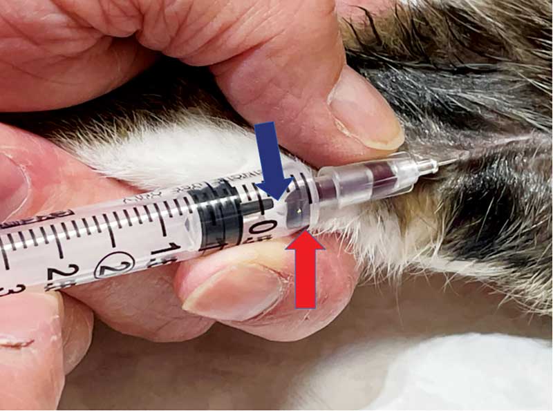
However, I am not using a tube like this because the amount of the vacuum often collapses the cat's small veins. Rather, I use a 3-cc syringe with a 25-gauge needle. Blood is drawn steadily and relatively slowly to avoid hemolysis of the sample. If a slow draw occurs, the speed of collection is further slowed.
Obstacle No. 2 and its solution. Because blood collection can be slow from the medial saphenous vein, clotting of the sample often occurs in the syringe, rendering the sample unacceptable for the chemistry machine and hematology machines.
The solution is the bead in the lithium heparin (LH) tube used with the chemistry machine. I take the bead out of the LH tube and put it in the syringe. (Figure 1) Although its stated purpose is to assist proper mixing of the blood sample, the bead has enough LH on it to anticoagulate the blood as soon as it enters the syringe. (Figure 2) With this approach, even the slowest of blood draws still yields an anticoagulated sample suitable for the chemistry machine.
Obstacle No. 3. Typically, the manufacturer's instructions state one should put 1.3 ml of blood in the LH tube to have the proper ratio between the blood and the LH; however, only about 0.25 ml are actually used to perform the test. Although some cats have medial saphenous veins that flow well, many do not. In some cats, collecting more than 0.5 ml of blood is not possible.
Obstacle No. 4. The manufacturer's instructions state only ethylenediamine tetraacetic acid (EDTA) treated blood is to be inserted into the hematology machine.
Solutions to No. 3 and No. 4. Blood is collected with a 3-cc syringe and 25-gauge needle. If I can get about 1.3-ml of blood, I do. If I can only get 0.5-ml, our study shows that is also acceptable.
The indicated rotors are loaded directly out of the syringe. The remainder of the blood is placed in the EDTA tube made for the hematology machine. The tube is placed in the machine, and the sample is run. Within about 15 minutes of blood collection, I have results of a chemistry profile with electrolytes, a total T4, and a CBC. I am now ready to talk to the owner about the cat's diagnosis and/or treatment plan.
Obstacle No. 5 and its solution. We have been taught heparin will change the morphology of the white blood cells, so blood smear interpretation will be inaccurate.
Six blood smears were made on the samples from each cat in the study. Three slides were made from blood immediately after the blood draw; it was not exposed to any anticoagulant. Three were made after being exposed to lithium heparin, plus EDTA. Each was stained with a Romanowsky-type rapid blood stain. They were read by a board-certified veterinary clinical pathologist. However, he was blinded as to the source of the blood smears. He was specifically tasked with observing for morphology changes that would make cell identification difficult. None were found, and all slides were deemed of sufficient quality for interpretation.
Overall outcome
If you do this, what is going to happen? First, you will use many more chemistry rotors and hematology reagents than in the past. Second, you will likely find the increase in volume will cause you to consider the purchase of one or more additional machines. (I have six chemistry machines and two hematology machines.) Third, your patient care will improve, because you are doing more blood-based diagnostics. Finally, your practice will grow as more and more satisfied clients tell their friends about the amazing care their cats received at your practice.
Note: This technique was validated for the Zoetis Diagnostics chemistry and hematology machines. Using it on other machines is not recommended until similar studies are performed on those machines.
Note: The only analyte that was adversely affected was the platelet count. It can be falsely increased or decreased. Therefore, if the platelet count does not seem valid, one should examine a stained blood smear.
Because of this technique, we perform blood tests on over 50 percent of the cats that we see. We average 337 chemistry profiles and 139 CBCs per month in our three-doctor feline practice. In addition, we also average 148 thyroid panels (TT4 + cholesterol). Imagine what this technique could do for your practice.
Gary D. Norsworthy, DVM, DABVP (feline), is the owner of Alamo Feline Health Center in San Antonio, Texas. He has been in private practice for 48 years, including 23 in feline-only practice. Dr. Norsworthy lectures frequently on feline diseases and is the editor and major author of seven feline textbooks. He is a board-certified feline specialist (one of only two in South Texas) and an adjunct professor at the College of Veterinary Medicine, Mississippi State University, and the Western University of Health Sciences. He received the 2020 Distinguished Career Achievement Award by the Texas Veterinary Medical Association.
Reference
1 Norsworthy, GD, Cook, AK, Lanier, CJ. Impact of preheparinization and sample volume on routine hematology findings in healthy cats. J Fel Med Surg. 2021;23(2); 79-85.
How Do You Draw Blood From a Cat
Source: https://www.veterinarypracticenews.com/challenges-in-feline-blood-collection/
0 Response to "How Do You Draw Blood From a Cat"
Post a Comment